| Citation: |
YAN Jun, DENG Xiaoqiong, HU Danjing, FANG Shibin, LIU Peijun, FANG Biao, HU Xianchao. Microscopic morphology of prismatic layers of freshwater cultured pearl[J]. South China Fisheries Science, 2013, 9(1): 48-52. DOI: 10.3969/j.issn.2095-0780.2013.01.008

|
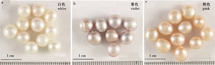
The microscopic morphology of the internal structure of freshwater cultured pearl with different colors were investigated by X-ray diffraction (XRD) and field emission-scanning electronic microscope (FE-SEM).Results clearly reveal that aragonite is found in high quality freshwater cultured pearls, but calcite and vaterite are not found, and the thickness of aragonite sheet in nacreous layer is not the same to pearls with different colors. The prismatic layer is near to the nucleus of the high quality pearls, which is made of a number of cylindrical or banded aragonite fibers. The fibers are oriented nearly parallel to the aragonite sheet. In addition, it is firstly observed that the aragonite fiber and sheet appear alternately in prismatic layer. Meanwhile, a modified model of the internal structure of freshwater cultured pearls is firstly established.
| [1] |
QIAO L, FENG Q L. Study on twin stacking faults in vaterite tablets of freshwater lacklustre pearls[J]. J Crystal Growth, 2007, 304(1): 253-256. doi: 10.1016/j.jcrysgro.2007.02.001
|
| [2] |
MA H Y, LEE L S. Characterization of vaternite in low quality freshwater-cultured pearls[J]. Mater Sci & Engin: C, 2006, 26(1): 721-723.
|
| [3] |
MA H Y, SU A A, ZHANG B L, et al. Vaterite or aragonite observed in the prismatic layer of freshwater-cultured pearls from South China[J]. Prog Nat Sci, 2009, 19(7): 817-820. doi: 10.1016/j.pnsc.2008.11.005
|
| [4] |
MA H Y, LI R K, YANG L X, et al. A modified integrated model of the internal structure of Chinese cultured pearls[J]. J Wuhan Univ Technol-Mater: Sci Ed, 2011, 26(3): 510-513. doi: 10.1007/s11595-011-0258-5
|
| [5] |
ZAREMBA C M, BELCHER A M, FRITZ M, et al. Critical transitions in the biofabrication of abalone shells and flat pearls[J]. Chem Mater, 1996, 8(3): 679-690. doi: 10.1021/cm9503285
|
| [6] |
WADA N, SUDA S, KANAMURA K, et al. Formation of thin calcium carbonate films with aragonite and vaterite forms coexisting with polyacrylic acids and chitosan membranes[J]. J Colloid & Interface Sci, 2004, 279(1): 167-174.
|
| [7] |
张刚生, 李浩璇. 淡水养殖珍珠的矿物组成特征[J]. 岩石矿物学杂志, 2004, 23 (1) : 89-93. https://xueshu.baidu.com/usercenter/paper/show?paperid=582d7941f98e2c722b872fe4bc85f9c8&site=xueshu_se&hitarticle=1
|
| [8] |
马红艳, 崔福斋. 淡水珍珠中球文石的XRD谱[J]. 矿物学报, 2003, 23(4): 371-373. https://xueshu.baidu.com/usercenter/paper/show?paperid=65d93a82f7aa460fd4b4e42b1376fd47&site=xueshu_se&hitarticle=1
|
| [9] |
秦作路, 马红艳, 木士春, 等. 优质淡水珍珠的体色及其与拉曼光谱的关系[J]. 矿物学报, 2007, 27(1): 73-76. https://xueshu.baidu.com/usercenter/paper/show?paperid=602a9bcc2f69fb491bfee4ea35d70b9c&site=xueshu_se&hitarticle=1
|
| [10] |
李耿, 林瓴, 沙拿利, 等. 淡水养殖珍珠的光泽颜色与有机质的关系初探[J]. 桂林工学院学报, 2007, 27(4): 569-571. https://xueshu.baidu.com/usercenter/paper/show?paperid=136p0mq0801q0cf09s720vf021660460&site=xueshu_se&hitarticle=1
|
| [11] |
MA Y F, GAO Y H, FENG Q L. Characterization of organic matrix extracted from freshwater pearls[J]. Mater Sci & Engin: C, 2011, 31(7): 1338-1342. https://xueshu.baidu.com/usercenter/paper/show?paperid=080cc0d1562f7da33f3d0ae7fd6bb65e&site=xueshu_se
|
| [12] |
孔蓓, 邹进福, 陈积光, 等. 海水养殖珍珠表层微形貌的结构研究——以广西防城养殖珍珠为例[J]. 矿产与地质, 2002, 16 (6): 341-345. https://xueshu.baidu.com/usercenter/paper/show?paperid=a92213fcac8b0b85f2b44b4c074a25b1&site=xueshu_se
|
| [13] |
曹莉嘉, 郭守国, 史凌云. 珍珠的光泽与其表面结构的关系研究[J]. 宝石和宝石学杂志, 2005, 7(3): 23-25. https://xueshu.baidu.com/usercenter/paper/show?paperid=57cd2d6f704c6a32c0887df756480005&site=xueshu_se&hitarticle=1
|
| [14] |
杜晓东, 邓陈茂. 珍珠的扫描电镜观察[J]. 湛江水产学院学报, 1991, 11(1): 10-15. https://xueshu.baidu.com/usercenter/paper/show?paperid=b380a3e262c37003fde864d4db95cfab&site=xueshu_se&hitarticle=1
|
| [15] |
谢玉坎. 珍珠科学[M]. 北京: 海洋出版社, 1995: 236.
|
| [16] |
张蓓丽. 系统宝石学[M]. 北京: 地质出版社, 1997: 390.
|
| [17] |
张妮, 郭继春, 张学云, 等. 珍珠表面形貌的AFM和SEM研究[J]. 岩石矿物学杂志, 2004, 24(4): 370-374. https://xueshu.baidu.com/usercenter/paper/show?paperid=3643747e7c5b49f1f7045bb73071eb21&site=xueshu_se&hitarticle=1
|
| [18] |
张妮, 郭继春, 张学云, 等. 淡水珍珠中文石球粒的发现与成珠机制探讨[J]. 矿物学报, 2005, 25(3): 307-311. https://xueshu.baidu.com/usercenter/paper/show?paperid=5019182b03feb62d245b7661d7c2061f&site=xueshu_se&hitarticle=1
|
| [19] |
张恩, 彭明生, 梁超伦, 等. 珍珠显微结构及纳米矿物的电镜研究[J]. 矿物学报, 2008, 28(2): 112-116.
|
| [20] |
马红艳, 韦起桂, 木士春, 等. 物理法改色前后优质淡水珍珠的超微结构特征研究[J]. 矿物学报, 2012, 32(1): 139-145. https://xueshu.baidu.com/usercenter/paper/show?paperid=968a793dbe0e430706e3c4f35c64908c&site=xueshu_se&hitarticle=1
|
| [21] |
张伟钢, 严俊, 汪港, 等. 一种天然湿敏性二维可调光子带隙材料及其光学性能研究[J]. 无机材料学, 2009, 24 (1): 57-60. https://xueshu.baidu.com/usercenter/paper/show?paperid=470d63e081559ecda4f0f0025cb128eb&site=xueshu_se&hitarticle=1
|
| [22] |
严俊, 张刚生. 褶纹冠蚌贝壳结构特征及其彩虹色呈色机制[J]. 安庆师范学院学报: 自然版, 2011, 17(3): 83-85. https://www.cnki.com.cn/Article/CJFDTOTAL-AQSX201103023.htm
|
| 1. |
张绍炜,戴小杰. 渔获量与自然死亡系数不确定性对印度洋黄鳍金枪鱼资源评估的影响. 海洋渔业. 2025(02): 141-152 .

| |
| 2. |
梁耀威,冯波,李美霖,孙钰,李忠炉. 基于多源渔获量数据的南海北部短尾大眼鲷可捕资源量评估. 中国水产科学. 2024(03): 332-342 .
| |
| 3. |
袁红春,史经伟. 基于改进YOLOv7- tiny和动态检测门的金枪鱼自动检测与计数研究. 渔业现代化. 2023(06): 74-83 .
| |
| 4. |
安康,官文江. 利用贝叶斯动态产量模型评估印度洋长鳍金枪鱼资源状态. 中国水产科学. 2023(09): 1142-1154 .
|
Supported by: Beijing Renhe Information Technology Co., Ltd.

 粤公网安备 44010502001741号

 DownLoad: